Fleurettes Retinoblastoma : Http Www Gmcjammu Nic In Retinoblastoma 20ppt Pdf - Necrosis is also very common and occurs when the tumor outgrows its vascular supply.
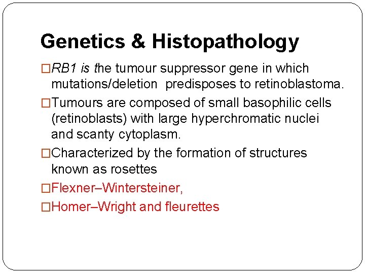
Fleurettes Retinoblastoma : Http Www Gmcjammu Nic In Retinoblastoma 20ppt Pdf - Necrosis is also very common and occurs when the tumor outgrows its vascular supply.. 10 many retinoblastomas have underlying elements of retinoma. Tumors composed entirely of fleurettes (retinoma/retinocytoma) are thought to be retinoblastoma precursors, and like retinoblastoma, harbor mutations in both copies of the rb1 gene. Calcification is almost pathognomonic of retinoblastoma, but its etiology is not known. American ophthalmology society first adopted the term retinoblastoma in 1926. • usually fatal due to meningeal spread, median survival of 9 months.
Necrosis is also very common and occurs when the tumor outgrows its vascular supply. Retinoblastoma is a major cancer treatment success story in developed countries where most deaths are caused by secondary tumors in germline mutation carriers. Incomplete shrinkage of the lesion may be noted after radiotherap). Our results show that s antigen may be a useful marker in the study of the embryologic development of the human. 10,31 comparison of adjacent normal retina, retinoma, and retinoblastoma showed loss of both rb1 alleles and early genomic copy number changes in retinoma that were amplified further in the adjacent retinoblastoma.
Pathology Of Retinoblastoma Pathology Made Simple from ilovepathology.com • primary retinoblastoma of pineal & parasellar sites. A tumor composed of fleurettes is deemed benign and called retinoma or retinocytoma. Incomplete shrinkage of the lesion may be noted after radiotherap). 10 many retinoblastomas have underlying elements of retinoma. Only one patient died (20 years after enucleation) because of metastatic osteosarcoma.in conclusion: 10,31 comparison of adjacent normal retina, retinoma, and retinoblastoma showed loss of both rb1 alleles and early genomic copy number changes in retinoma that were amplified further in the adjacent retinoblastoma. Fleurettes are considered the most differentiated form of rosette found in the tumor. Necrosis is also very common and occurs when the tumor outgrows its vascular supply.
Calcification is almost pathognomonic of retinoblastoma, but its etiology is not known.
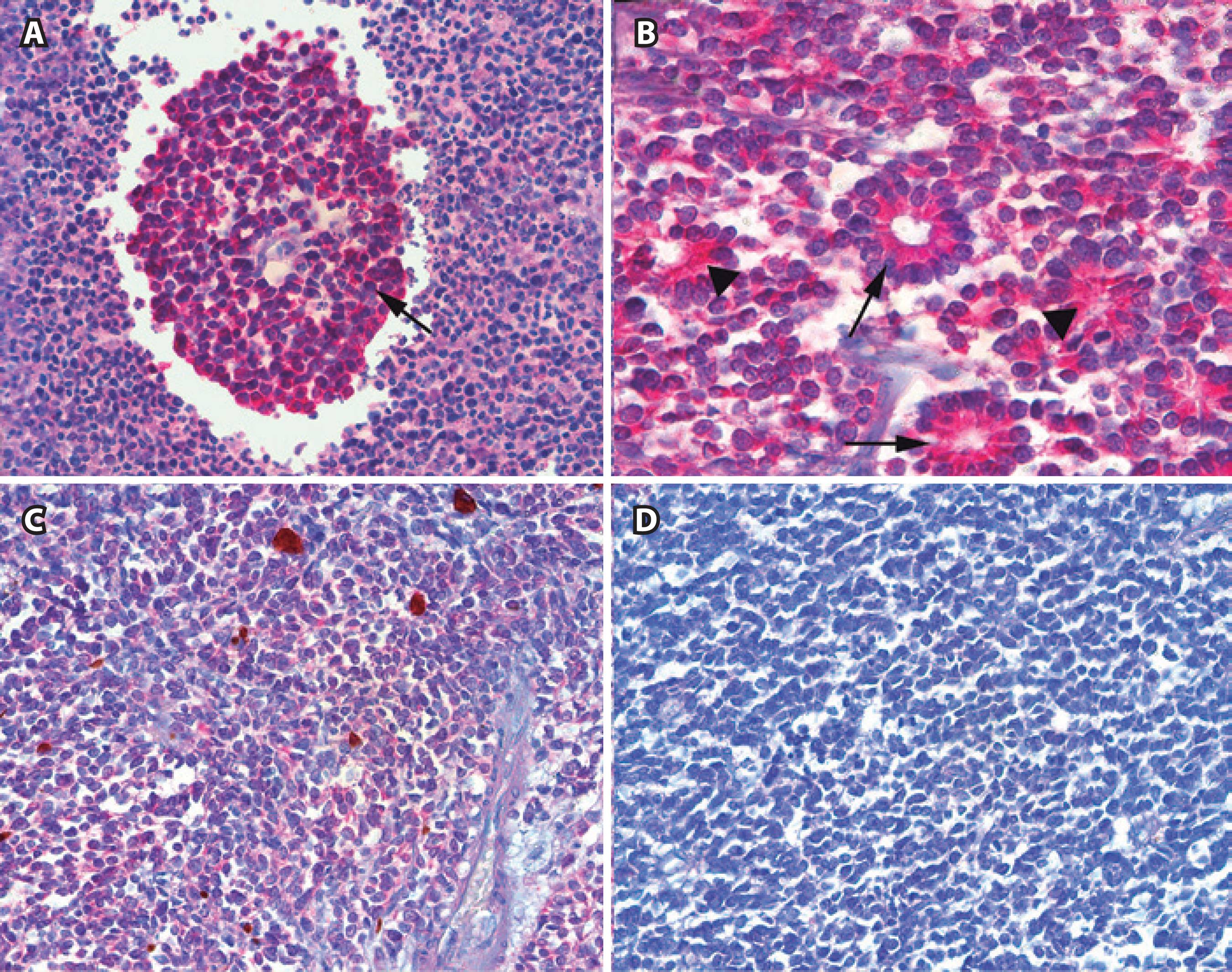
10,31 comparison of adjacent normal retina, retinoma, and retinoblastoma showed loss of both rb1 alleles and early genomic copy number changes in retinoma that were amplified further in the adjacent retinoblastoma. Retinoblastoma is a rare cancer of the infant retina that is diagnosed in approximately 8,000 children each year worldwide. True spontaneous regression of retinoblastoma is rare, but is probably due to extensive tumor necrosis and central retinal artery occlusion, resulting in phthisis bulbi. Most common intraocular tumor of children with incidence of 1 per 20,000 live births. Fleurettes (figure 3c) are retinoblastoma cells that have undergone greater photoreceptor differentiation and group together as a bouquet. Incomplete shrinkage of the lesion may be noted after radiotherap). Fleurettes are retinoblastoma cells that have undergone greater photoreceptor differentiation. A tumor composed of fleurettes is deemed benign and called retinoma or retinocytoma. Pathology of retinoma reveals nonproliferative fleurettes. Fleurettes are a higher form of photoreceptor differentiation than rosettes. May be congenital but not recognized until ages 6 months to 2 years. 15% to 20% of retinoblastoma as harbor very well differentiated foci of actual photoreceptor differentiation. Only one patient died (20 years after enucleation) because of metastatic osteosarcoma.in conclusion:
Retinoblastoma histopathology is a combination of undifferentiated cells and areas of tumor differentiation shown as rosettes and fleurettes. Fleurettes are a higher form of photoreceptor differentiation than rosettes. Retinoblastoma is considered to be one of the types of embryonal central nervous system tumors, a group which includes medulloblastoma, neuroblastoma, pineoblastoma, and medulloepithelioma, among others. Retinoma discovered in a retinoblastoma eye removed after the main active tumor dispersed throughout the vitreous following 1 cycle of chemotherapy. 10 many retinoblastomas have underlying elements of retinoma.
Scielo Brasil Immunohistochemical Analysis Of Retinoblastoma Cell Phenotype Using Neuronal And Glial Cell Markers Immunohistochemical Analysis Of Retinoblastoma Cell Phenotype Using Neuronal And Glial Cell Markers from minio.scielo.br Necrosis is also very common and occurs when the tumor outgrows its vascular supply. Necrotic cells appear pink on h&e staining. Calcification is almost pathognomonic of retinoblastoma, but its etiology is not known. Tumors composed entirely of fleurettes (retinoma/retinocytoma) are thought to be retinoblastoma precursors, and like retinoblastoma, harbor mutations in both copies of the rb1 gene. Fleurettes are a higher form of photoreceptor differentiation than rosettes. 10 many retinoblastomas have underlying elements of retinoma. Retinoblastoma is a major cancer treatment success story in developed countries where most deaths are caused by secondary tumors in germline mutation carriers. Traditionally, level of neoplastic differentiation is a crucial index of adjuvant chemotherapy and prognosis.
10 many retinoblastomas have underlying elements of retinoma.
10 many retinoblastomas have underlying elements of retinoma. American ophthalmology society first adopted the term retinoblastoma in 1926. Retinoblastoma • ophthalmic cap approved * data elements with asterisks are not required for accreditation purposes for the commission on cancer. Only one patient died (20 years after enucleation) because of metastatic osteosarcoma.in conclusion: Traditionally, level of neoplastic differentiation is a crucial index of adjuvant chemotherapy and prognosis. Retinoblastoma histopathology is a combination of undifferentiated cells and areas of tumor differentiation shown as rosettes and fleurettes. Necrosis is also very common and occurs when the tumor outgrows its vascular supply. Incomplete shrinkage of the lesion may be noted after radiotherap). Fleurettes are a higher form of photoreceptor differentiation than rosettes. No other retinal cell types were found. Tumors composed entirely of fleurettes (retinoma/retinocytoma) are thought to be retinoblastoma precursors, and like retinoblastoma, harbor mutations in both copies of the rb1 gene. Calcification is almost pathognomonic of retinoblastoma, but its etiology is not known. • primary retinoblastoma of pineal & parasellar sites.
Pathology of retinoma reveals nonproliferative fleurettes. Necrosis is also very common and occurs when the tumor outgrows its vascular supply. Need mutations in both alleles to inactivate rb gene, a. May be congenital but not recognized until ages 6 months to 2 years. Fleurettes (figure 3c) are retinoblastoma cells that have undergone greater photoreceptor differentiation and group together as a bouquet.
Retinoblastoma Dr Anupam Assistant Professor Ophthalmology Epidemiology The from slidetodoc.com True spontaneous regression of retinoblastoma is rare, but is probably due to extensive tumor necrosis and central retinal artery occlusion, resulting in phthisis bulbi. 15% to 20% of retinoblastoma as harbor very well differentiated foci of actual photoreceptor differentiation. Retinoblastoma histopathology is a combination of undifferentiated cells and areas of tumor differentiation shown as rosettes and fleurettes. Our results show that s antigen may be a useful marker in the study of the embryologic development of the human. Fleurettes are a higher form of photoreceptor differentiation than rosettes. Calcification is common in these tumors. This comparative study showed that rosettes and fleurettes of retinoblastoma are an attempt to differentiate photoreceptor cells. American ophthalmology society first adopted the term retinoblastoma in 1926.
Incomplete shrinkage of the lesion may be noted after radiotherap).
A tumor composed of fleurettes is deemed benign and called retinoma or retinocytoma. Fleurettes (figure 3c) are retinoblastoma cells that have undergone greater photoreceptor differentiation and group together as a bouquet. True spontaneous regression of retinoblastoma is rare, but is probably due to extensive tumor necrosis and central retinal artery occlusion, resulting in phthisis bulbi. Necrosis is also very common and occurs when the tumor outgrows its vascular supply. Fleurettes are considered the most differentiated form of rosette found in the tumor. Fleurettes lack mitosis or necrosis. 10 many retinoblastomas have underlying elements of retinoma. The degree of differentiation in retinoblastoma is determined by the development of rosettes and fleurettes, which has been confirmed by electron microscopy and tissue culture. Retinoblastoma histopathology is a combination of undifferentiated cells and areas of tumor differentiation shown as rosettes and fleurettes. These elements may be clinically important, but are not yet validated or regularly used in patient management. Retinoma histology showing abundant fleurettes and sparse cells with eosinophilic cytoplasm. Retinoma discovered in a retinoblastoma eye removed after the main active tumor dispersed throughout the vitreous following 1 cycle of chemotherapy. • primary retinoblastoma of pineal & parasellar sites.
Tumors composed entirely of fleurettes (retinoma/retinocytoma) are thought to be retinoblastoma precursors, and like retinoblastoma, harbor mutations in both copies of the rb1 gene fleuret. Retinoma histology showing abundant fleurettes and sparse cells with eosinophilic cytoplasm.


Posting Komentar
0 Komentar